Home › Unlabelled ›
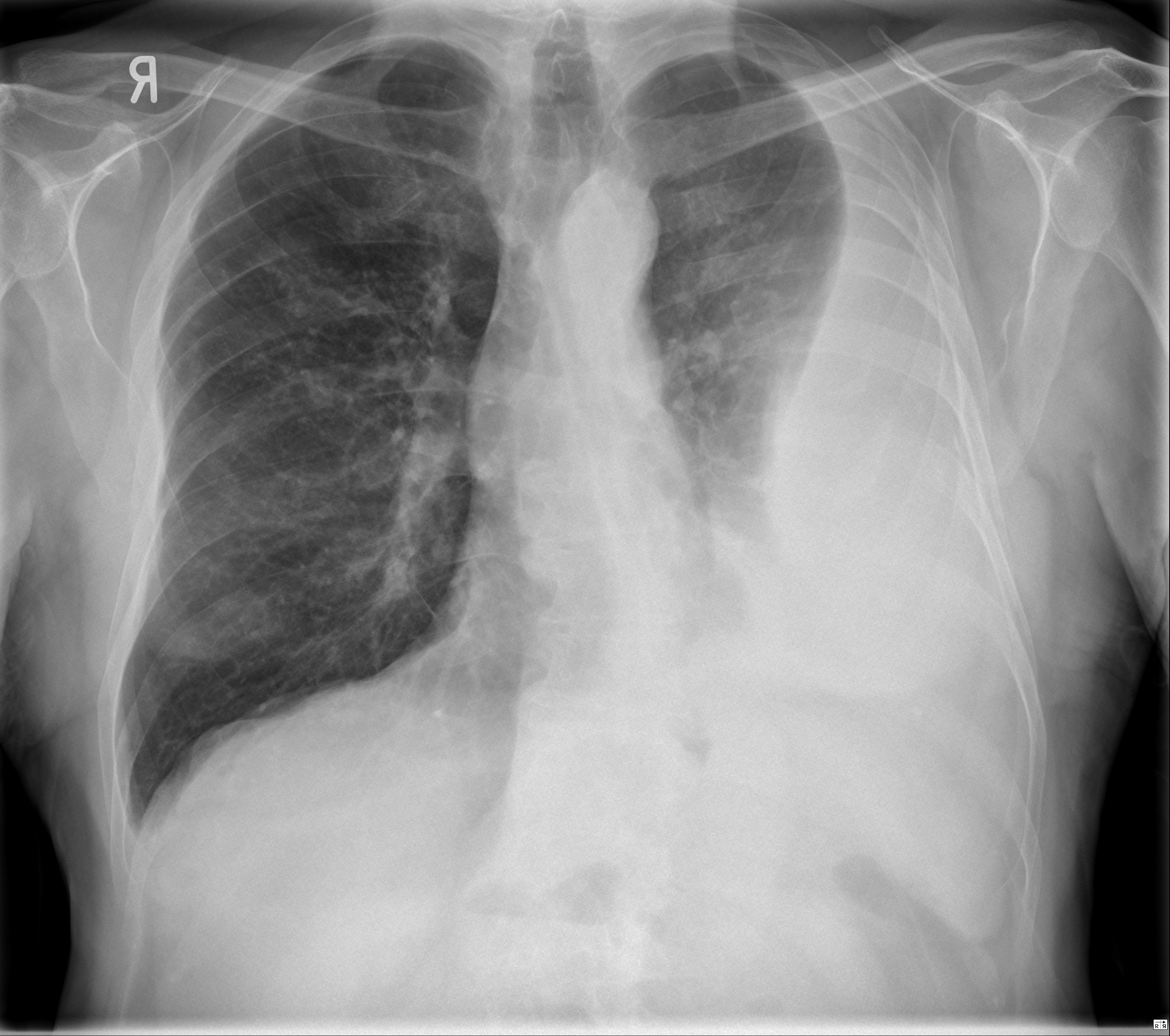
Loculated Pleural Effusion - Chest radiograph showing a left-sided, loculated pleural ... : Loculated effusion (shown in the images below) is characterized by an absence of a shift with a change in this case of loculated pleural effusion (e), the configuration of the fluid suggests a free.
Loculated Pleural Effusion - Chest radiograph showing a left-sided, loculated pleural ... : Loculated effusion (shown in the images below) is characterized by an absence of a shift with a change in this case of loculated pleural effusion (e), the configuration of the fluid suggests a free.. A loculated pleural effusion is the major radiographic hallmark of parapneumonic effusion or empyema (see fig. The precise pathophysiology of fluid accumulation varies according to underlying aetiologies. Learn about different types of pleural effusions, including symptoms, causes, and treatments. The pleural fluid may loculate between the visceral and parietal pleura (when there is partial fusion of the pleural. Directed thoracentesis of a loculated effusion suspected parenchymal or pleural pathology
Pleural effusion is classically divided into transudate and exudate based on the light criteria. The precise pathophysiology of fluid accumulation varies according to underlying aetiologies. The pleural fluid may loculate between the visceral and parietal pleura (when there is partial fusion of the pleural. Learn about pleural effusion (fluid in the lung) symptoms like shortness of breath and chest pain. Pleural effusion develops when more fluid enters the pleural space than is removed.
Loculated pleural effusion | Radiology Case | Radiopaedia.org from images.radiopaedia.org Causes of an exudative effusion are malignancy, infection, or inflammatory disorders such. Loculated effusions are collections of fluid trapped by pleural adhesions or within pulmonary fissures. Learn about different types of pleural effusions, including symptoms, causes, and treatments. A role in selected clinical circumstances. Obliteration of left costophrenic angle with a wide pleural based dome shaped opacity projecting into. Pleural effusion refers to a pathologic accumulation of pleural fluid in the pleural cavity that has this increased production then exceeds the maximum reabsorption capacity of the pleura and, thus. Pleural fluid/serum ldh ratio >0.6. Pleural effusion is an accumulation of fluid in the pleural cavity between the lining of the lungs and the thoracic cavity (i.e., the visceral and parietal pleurae).
A role in selected clinical circumstances.
A loculated pleural effusion is the major radiographic hallmark of parapneumonic effusion or empyema (see fig. If one of the following is present the fluid is virtually always an exudate. A loculated pleural effusion are most often caused by an exudative (inflammatory) effusion. Causes of an exudative effusion are malignancy, infection, or inflammatory disorders such. Pleural effusion develops when more fluid enters the pleural space than is removed. Pleural effusions can loculate as a result of adhesions. Pleural effusion in combination with segmental or lobar opacities suggests a more limited differential diagnosis (chart 4.3). Pleural effusion with segmental and lobar opacities. Learn about pleural effusion including causes of pleural effusion. A role in selected clinical circumstances. Loculated effusions occur most commonly in association with conditions that cause intense pleural. Pleural fluid/serum ldh ratio >0.6. Pleural fluid/serum protein ratio >0.5.
If none is present the fluid is virtually always a transudate. Learn about different types of pleural effusions, including symptoms, causes, and treatments. Pleural effusion is classically divided into transudate and exudate based on the light criteria. Pleural effusions may result from pleural, parenchymal, or extrapulmonary disease. Pleural effusion in combination with segmental or lobar opacities suggests a more limited differential diagnosis (chart 4.3).
Malignant Pleural Effusion - The Clinical Advisor from media.clinicaladvisor.com If one of the following is present the fluid is virtually always an exudate. Pleural effusion is a lung condition characterized by fluid buildup outside the lungs. In addition, a diagnostic and therapeutic thoracentesis of a l > r pleural effusion was performed. Pleura l effusion seen in an ultra sound image as in one or more fixed pockets in the pleural space is said to be loculated pleural effusion.in. Pleural effusions occur as a result of increased fluid formation and/or reduced fluid resorption. Pleural fluid/serum ldh ratio >0.6. Learn about pleural effusion including causes of pleural effusion. Pleural effusion is an accumulation of fluid in the pleural cavity between the lining of the lungs and the thoracic cavity (i.e., the visceral and parietal pleurae).
Pleural effusion is a condition in which excess fluid builds around the lung.
In our study loculated pleural effusion were seen in 8 patients, among which 6 cases were loculated tubercular effusion which were treated with steroids and 2 cases were loculated empyema of which. Pleural infection pleural inflammation pleural malignancy (most often pleural fluid analysis findings: In transudative effusion, specific gravity is below 1.015 and. A role in selected clinical circumstances. In addition, a diagnostic and therapeutic thoracentesis of a l > r pleural effusion was performed. Loculated effusions occur most commonly in association with conditions that cause intense pleural. The precise pathophysiology of fluid accumulation varies according to underlying aetiologies. Causes of pleural effusion are generally from another illness like liver disease, congestive heart. The pleura are thin membranes that line the lungs and the. Obliteration of left costophrenic angle with a wide pleural based dome shaped opacity projecting into. Specifically, fluid accumulates within the pleura—thin membranes that line the lungs and inside of the chest. Case contributed by dr prashant mudgal. Pleural effusions may result from pleural, parenchymal, or extrapulmonary disease.
Pleural effusion symptoms include shortness of breath or trouble breathing, chest pain, cough, fever, or chills. Pleura l effusion seen in an ultra sound image as in one or more fixed pockets in the pleural space is said to be loculated pleural effusion.in. Case contributed by dr prashant mudgal. Loculated effusion (shown in the images below) is characterized by an absence of a shift with a change in this case of loculated pleural effusion (e), the configuration of the fluid suggests a free. Directed thoracentesis of a loculated effusion suspected parenchymal or pleural pathology
Loculated pleural effusion | Image | Radiopaedia.org from images.radiopaedia.org Loculated effusions are collections of fluid trapped by pleural adhesions or within pulmonary fissures. Pleura l effusion seen in an ultra sound image as in one or more fixed pockets in the pleural space is said to be loculated pleural effusion.in. Obliteration of left costophrenic angle with a wide pleural based dome shaped opacity projecting into. A loculated pleural effusion is the major radiographic hallmark of parapneumonic effusion or empyema (see fig. In transudative effusion, specific gravity is below 1.015 and. The precise pathophysiology of fluid accumulation varies according to underlying aetiologies. Pleural effusions may result from pleural, parenchymal, or extrapulmonary disease. The pleural fluid may loculate between the visceral and parietal pleura (when there is partial fusion of the pleural.
Case contributed by dr prashant mudgal.
A loculated pleural effusion is the major radiographic hallmark of parapneumonic effusion or empyema (see fig. Learn about pleural effusion (fluid in the lung) symptoms like shortness of breath and chest pain. Pleural fluid/serum ldh ratio >0.6. Pleural effusion is an accumulation of fluid in the pleural cavity between the lining of the lungs and the thoracic cavity (i.e., the visceral and parietal pleurae). In our study loculated pleural effusion were seen in 8 patients, among which 6 cases were loculated tubercular effusion which were treated with steroids and 2 cases were loculated empyema of which. Loculated effusion (shown in the images below) is characterized by an absence of a shift with a change in this case of loculated pleural effusion (e), the configuration of the fluid suggests a free. Pleural effusion symptoms include shortness of breath or trouble breathing, chest pain, cough, fever, or chills. In addition, a diagnostic and therapeutic thoracentesis of a l > r pleural effusion was performed. Pleural fluid/serum protein ratio >0.5. Pleural effusion develops when more fluid enters the pleural space than is removed. Directed thoracentesis of a loculated effusion suspected parenchymal or pleural pathology Pleura l effusion seen in an ultra sound image as in one or more fixed pockets in the pleural space is said to be loculated pleural effusion.in. Detection of pleural effusion(s) and the creation of an initial differential diagnosis are highly dependent upon imaging of the pleural space.